
Clinical Case
Aesthetic Smile Rehabilitation Using IPS e.max MT Partial Crowns
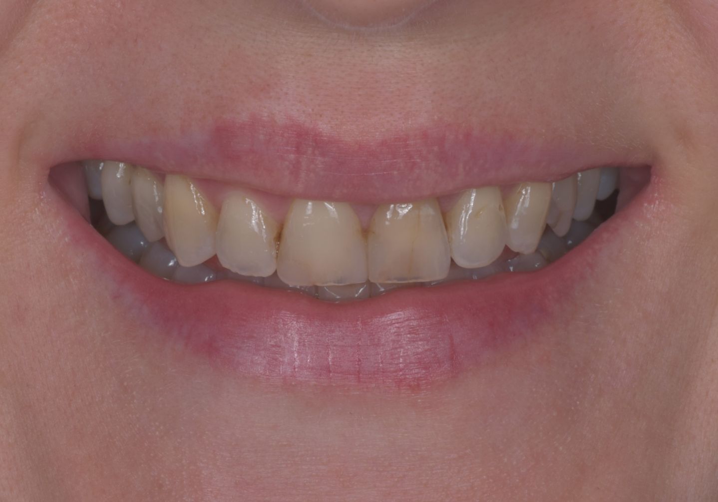

Initial Condition and Treatment Plan
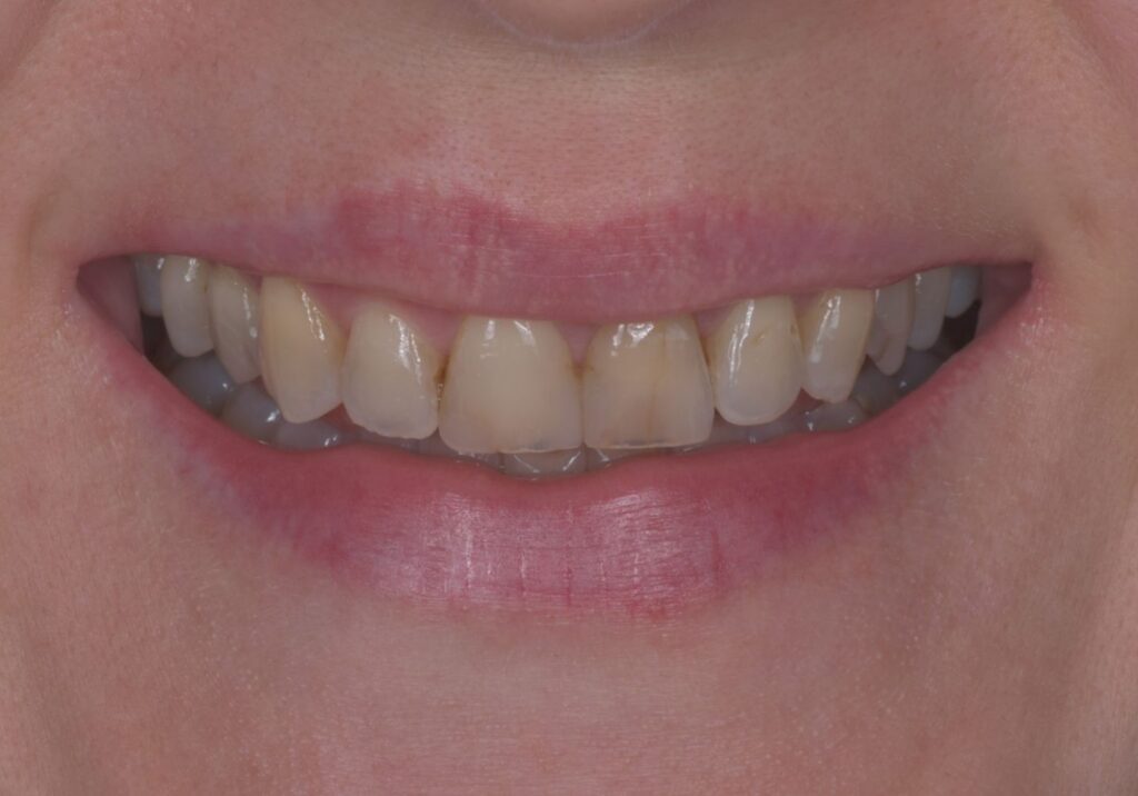
The patient visited our clinic seeking to improve the aesthetics of her smile. Her primary concern was the color of her front teeth, which appeared dark and uneven. She was particularly bothered by a root canal–treated front tooth that had become significantly darker than the adjacent teeth, disrupting the overall harmony of her smile.
During the clinical examination, it was also found that the posterior premolars and molars had been extensively restored with older composite fillings that offered a limited long-term prognosis. In addition, several teeth had lost their natural anatomy, and the overall dentition exhibited noticeable color inconsistency.
The initial diagnostic assessment revealed that the palatal (inner) walls of the upper anterior teeth had been significantly reduced and weakened. As a result, conventional veneers were not a suitable treatment option. Instead, all-ceramic partial crowns were indicated, allowing the natural tooth anatomy to be restored while providing sufficient structural strength and achieving an optimal aesthetic result.
Following a comprehensive diagnostic evaluation, a complete aesthetic rehabilitation of the upper arch using all-ceramic restorations was planned. The treatment plan included replacing the existing defective restorations, digitally designing the new smile, and fabricating a total of 12 all-ceramic partial crowns. Both the anterior and posterior teeth were restored with all-ceramic partial crowns.
The goal of the treatment was to create a natural, harmonious smile while preserving as much healthy hard dental tissue as possible.
Treatment plan proposal
Treatment plan
Phase 1 – Diagnostics (1 month before treatment)
- Clinical examination
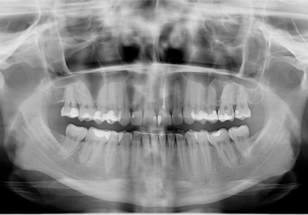
- OPG, CBCT, photographic documentation and digital intraoral scan
- Diagnosis
- Treatment plan
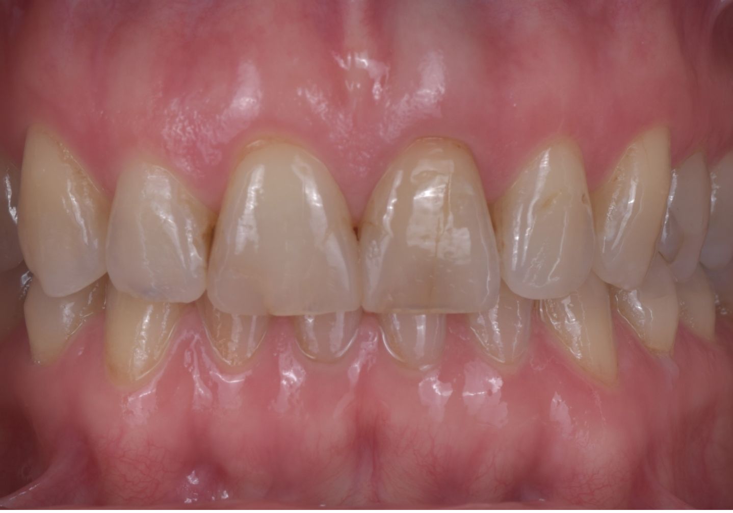


Phase 2 – Day 1 of treatment – Pre-prosthetic preparation and digital planning
- Removal of old composite fillings
- Reconstruction of the teeth with new composite fillings
- Digital Smile Design
- Digital CAD/CAM planning
- Digital wax-up
- Design of the new tooth anatomy, length, and shape
- Fabrication of diagnostic models in our in-house laboratory
- Fabrication of a silicone index (mock-up)
- Laboratory work: approximately 6 hours
Phase 3 – Day 2 of treatment – Mock-up and preparation of the anterior teeth
- Transfer of the mock-up to the patient's mouth
- Approval of the tooth anatomy, length, and shade
- Preparation of 6 anterior teeth
- Digital intraoral scan
- Fabrication and placement of temporary restorations
Phase 4 – Day 3 of treatment – Preparation of the posterior teeth
- Preparation of 6 posterior teeth
- Final digital impressions
- Design of ceramic restorations in CAD software
- Milling of IPS e.max MT partial crowns
- Characterization and glazing of ceramic restorations
- Laboratory work: approximately 6 hours
Phase 5 – Day 4 of treatment – Adhesive cementation
- Try-in of all restorations
- Evaluation of shade, anatomy, and contacts
- Adhesive cementation of partial crowns
- Occlusion check
- Bite adjustment
- Photographic documentation
- Home oral hygiene instructions
- OPG

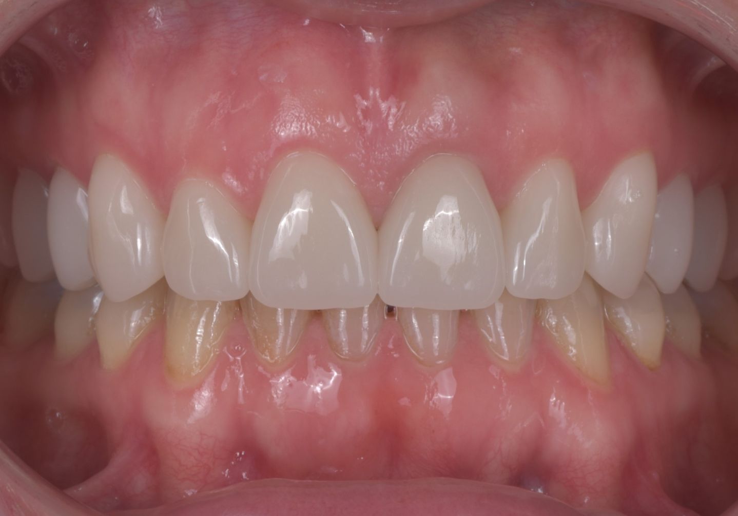
Phase 6 – Follow-up visit
- Soft tissue evaluation
- Evaluation of ceramic restorations
- Occlusion check
- Minor bite adjustment
- Final photographic documentation
ALL ON 4
Treatment process
Phase 1 – Diagnostics (2 hours, 1 month before treatment)
Approximately one month before the start of treatment, the patient underwent an initial consultation and complete digital diagnostics.
The examination included:
- clinical examination,
- panoramic X-ray (OPG),
- CBCT (3D CT),
- professional photographic documentation,
- digital intraoral scan.
Based on the collected data, an analysis of smile aesthetics, tooth anatomy, occlusion, and the condition of existing restorations was performed. After a consultation with the patient, an individual treatment plan was created, and digital planning of the new reconstruction was initiated.
Phase 2 – Day 1 of treatment – Pre-prosthetic preparation
On the first day of treatment, all unsatisfactory composite fillings were removed and replaced with new restorations, creating a stable foundation for the subsequent prosthetic treatment.
After completing this phase, digital planning of the new smile was carried out.
Digital treatment planning (laboratory phase – 6 hours)
In specialized CAD/CAM software, data from CBCT, digital intraoral scans, photographic documentation, and analysis of intermaxillary relationships were combined. Based on this data, a virtual reconstruction of the future dentition was created, including the new tooth anatomy, length, shape, aesthetic smile line, and final shade.
In our in-house CAD/CAM laboratory, a digital wax-up and diagnostic models of the new reconstruction were subsequently fabricated. The entire laboratory phase of digital planning took approximately 6 hours.
Phase 3 – Day 2 of treatment – Mock-up and preparation of the anterior teeth (4 hours)
Based on the digital wax-up, a mock-up was created using a silicone index and transferred directly into the patient’s mouth.
Before the preparation, the patient was able to try the new shape, length, anatomy, and aesthetics of her future smile. After a joint consultation, she approved the final anatomy and shade of the future ceramic restorations.
Subsequently, a minimally invasive preparation of six anterior teeth was performed precisely according to the approved mock-up. This approach ensured that only the necessary amount of healthy hard dental tissue was removed.
After completing the preparation, a digital intraoral scan was performed and temporary restorations were fabricated.
Phase 4 – Day 3 of treatment – Preparation of the posterior teeth (3–4 hours)
During the third visit, six posterior teeth intended for all-ceramic partial crowns were prepared.
After completing the preparation, final digital intraoral impressions of all prepared teeth were taken.
Laboratory fabrication of ceramic restorations (6 hours)
Based on the digital impressions, the fabrication of the definitive ceramic restorations was started in our in-house CAD/CAM laboratory.
The following restorations were fabricated:
- ceramic partial crowns,
- an all-ceramic crown,
- ceramic partial crowns.
The restorations were made from IPS e.max MT material in shade A1. After milling, they were individually characterized, glazed, and prepared for adhesive cementation.
The total laboratory fabrication time was approximately 6 hours.
Phase 5 – Day 4 of treatment – Adhesive cementation of ceramic restorations (4 hours)
During the final visit, all ceramic restorations were first tried in. The dentist checked the accuracy of their fit, anatomy, contact points, shade, and occlusion.
After the patient approved the result, adhesive cementation of the ceramic partial crowns was performed.
After completing the cementation, the following procedures were carried out:
- occlusion check,
- minor bite adjustment,
- final photographic documentation,
- evaluation of the aesthetics and function of the entire reconstruction,
- home oral hygiene instructions.
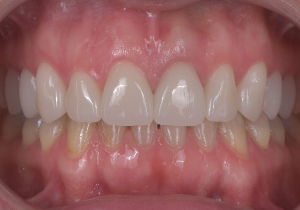
The patient was very satisfied with the treatment outcome and especially appreciated the natural tooth anatomy, their length, shape, unified shade, and the overall harmony of her new smile.
Phase 6 – Follow-up visit after cementation (30 minutes)
During the follow-up visit, the ceramic restorations, soft tissues, and occlusion were evaluated.
A minor bite adjustment was performed if needed, final photographic documentation was taken, and the patient’s adaptation to the new restorations was assessed.
The patient experienced no difficulties with speaking or chewing. The restorations were fully functional, aesthetically harmonious, and the patient was extremely satisfied with the treatment outcome.
Treatment outcome
The aesthetic rehabilitation was completed within 4 days of active treatment, with the initial diagnostics performed 1 month before the start of treatment.
The treatment achieved:
- unification of the shade of the entire dentition,
- elimination of the aesthetic discrepancy caused by the endodontically treated tooth,
- creation of new tooth anatomy,
- restoration of proper proportions and incisal length,
- more natural smile aesthetics,
- replacement of unsatisfactory fillings with all-ceramic restorations,
- a long-term stable and aesthetic result.
Treatment methods used
- OPG
- CBCT (3D CT)
- 3Shape digital intraoral scanner
- Digital Smile Design
- Digital CAD/CAM planning
- Digital wax-up
- Silicone mock-up
- CAD/CAM laboratory
- IPS e.max MT
- Collaboration with a specialized dental technician

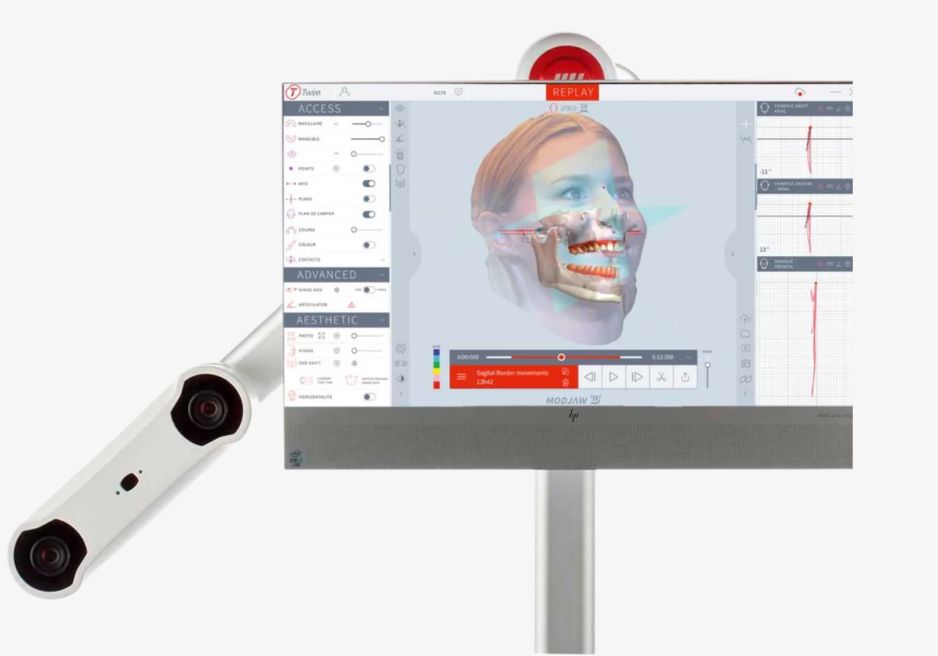


Overall Assessment
The patient expressed complete satisfaction with the treatment outcome. She particularly appreciated the natural appearance of her new smile, the harmonious tooth anatomy, the shape and length of her teeth, and the uniform A1 shade, which fully met her aesthetic expectations.



Appointment available within 24 hours
We know you don’t want to wait when it comes to your health. Fast scheduling for an examination and treatment within 2 business days.
Everything under one roof.
You don’t need to go anywhere else — we handle everything in one place, with a team of experienced specialists who work closely together.
High-quality materials with a warranty.
The highest-quality biocompatible materials ensure long-lasting restorations and comfort during everyday use.
Precise 3D diagnostics.
Thanks to advanced 3D technology, we can see every detail. This allows us to plan treatment with maximum precision and safety.
“Dental health is not only about technology – it is above all about trust, empathy, and cooperation between the doctor and the patient.”
satisfied patients
years of experience in dentistry
beautiful smiles
certificates and qualifications
Your journey to the perfect smile starts here
Book your consultation today!
Take the first step towards your dream smile – book your consultation today!