
Clinical Case
All-on-4
Full-Mouth Rehabilitation Using the All-on-4® Concept with Immediate Implant Loading and Definitive Zirconia Prostheses on Individual Titanium Bars
Initial Condition and Treatment Plan
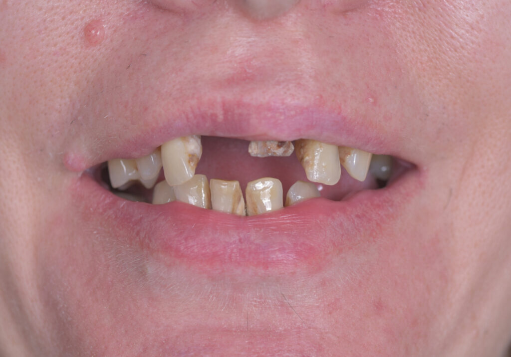
The patient presented to our clinic seeking a comprehensive solution for a long-standing, severely compromised dentition that had significantly affected not only the aesthetics of her smile but also masticatory function, eating comfort, and overall quality of life.
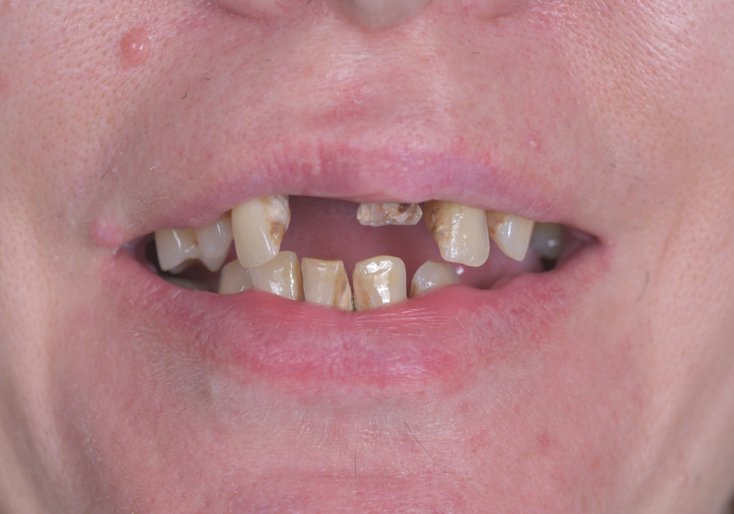
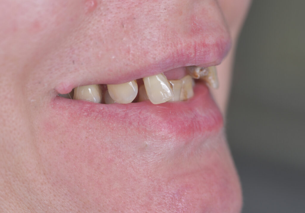
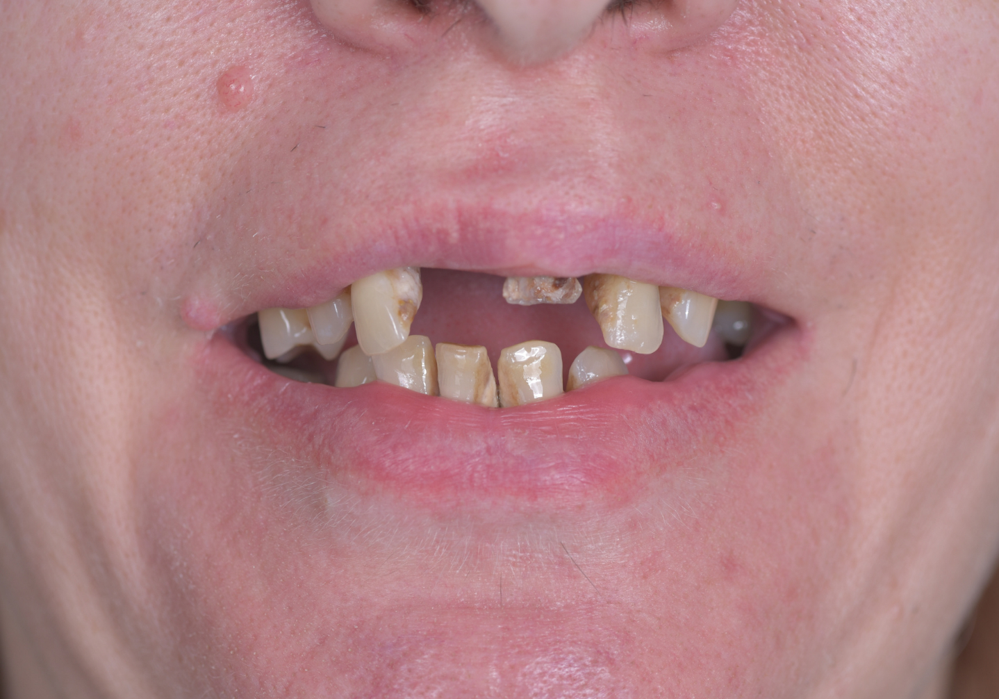
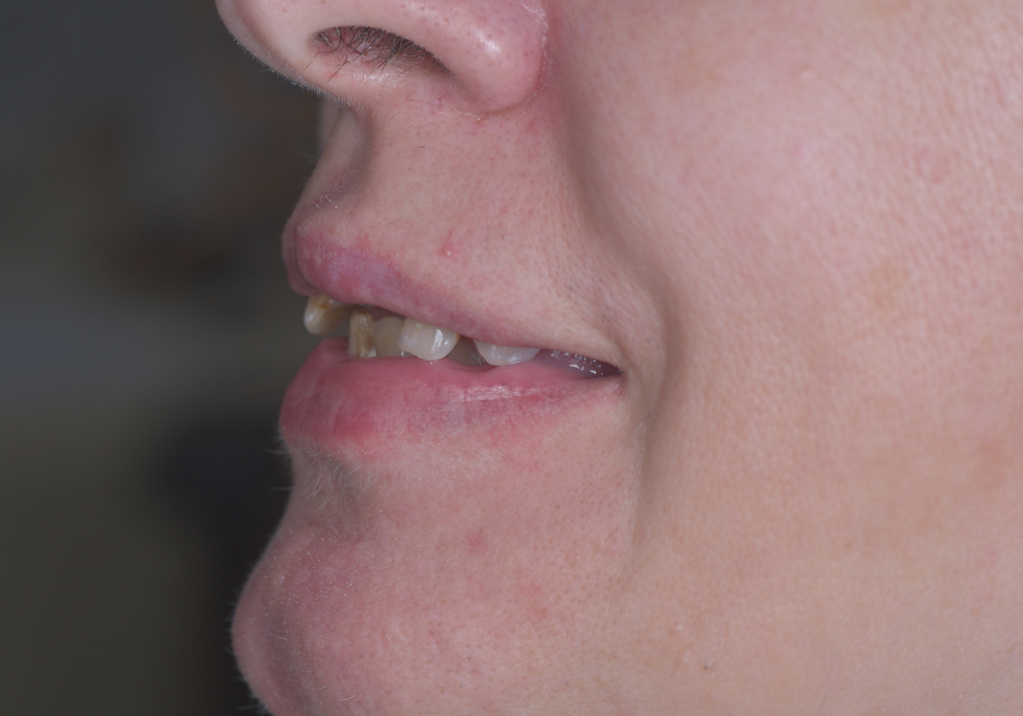
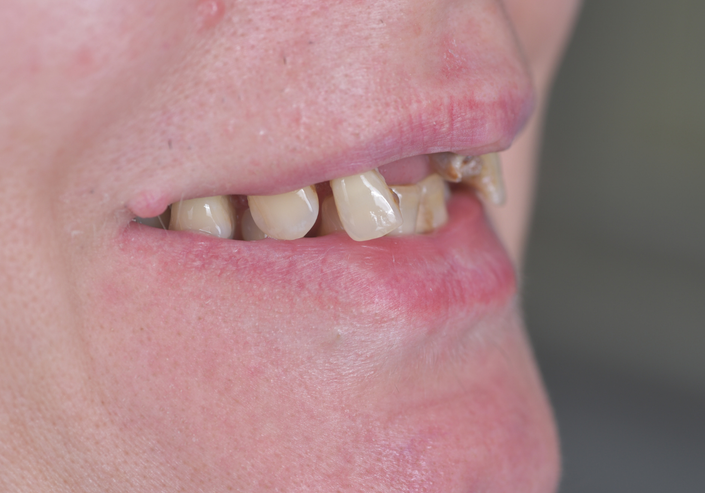
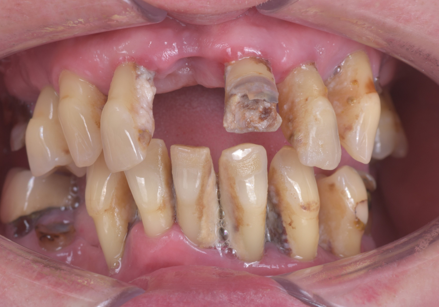
The initial clinical examination revealed generalized periodontitis affecting the entire dentition. Clinical findings included extensive gingival inflammation, multiple carious lesions, several retained root fragments (radices), and Grade III pathological mobility of all remaining teeth. The long-term prognosis for preserving the natural dentition was considered hopeless.
In addition, the patient exhibited severe malocclusion, disturbed intermaxillary relationships, and the absence of several posterior teeth. These conditions resulted in uneven distribution of occlusal forces, overloading of the remaining teeth, progressive collapse of the occlusion, and deterioration of facial aesthetics.
Following comprehensive clinical and radiographic evaluation, a treatment plan was established for complete oral rehabilitation using the All-on-4® concept in both the maxilla and the mandible, including immediate implant loading and subsequent fabrication of definitive screw-retained zirconia prostheses supported by individually milled titanium bars.
Clinical Case
Initial Clinical Findings



Diagnostics
Treatment Plan
Phase 1 – Diagnostic Examination
- Comprehensive clinical examination, Panoramic radiograph (OPG), Cone Beam Computed Tomography (CBCT)
- Clinical photography
- Digital intraoral scanning
- Diagnosis and preparation of an individualized treatment plan

Phase 2 – Preoperative Preparation
- Professional deep dental cleaning
- Soft tissue laser therapy
- Atraumatic extraction of retained roots (teeth #16, #24, and #25)
- New digital intraoral scan following preoperative treatment
- Oral hygiene instruction and patient education

Digital Treatment Planning
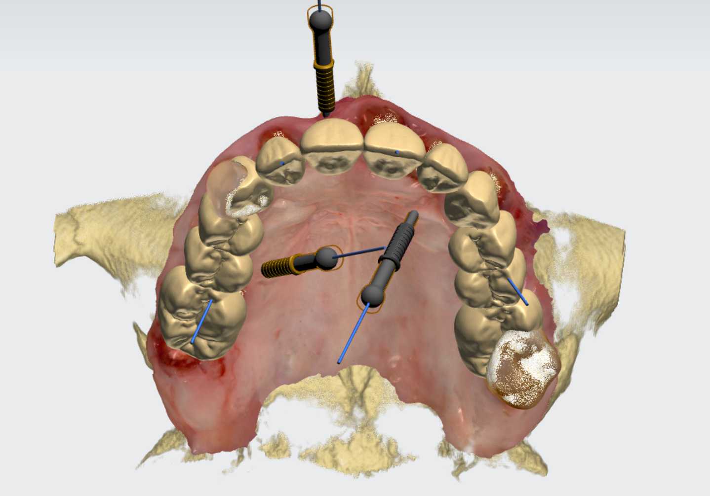
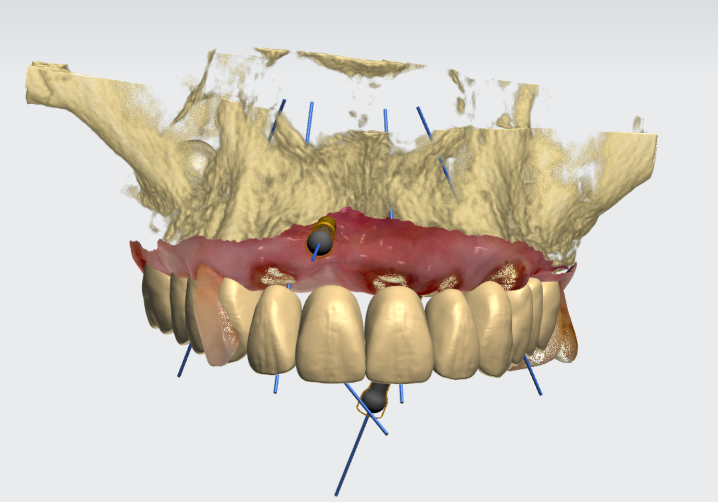
- Digital design of the new occlusion, tooth anatomy, and smile aesthetics
- Fabrication of digital and physical models of both dental arches
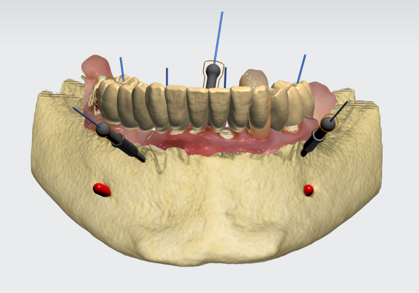
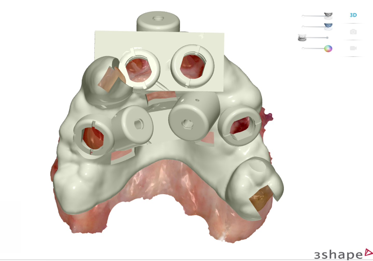
- Manufacturing of two fully guided surgical templates for the maxilla and mandible
- Manufacturing of Lower Surgical Guides
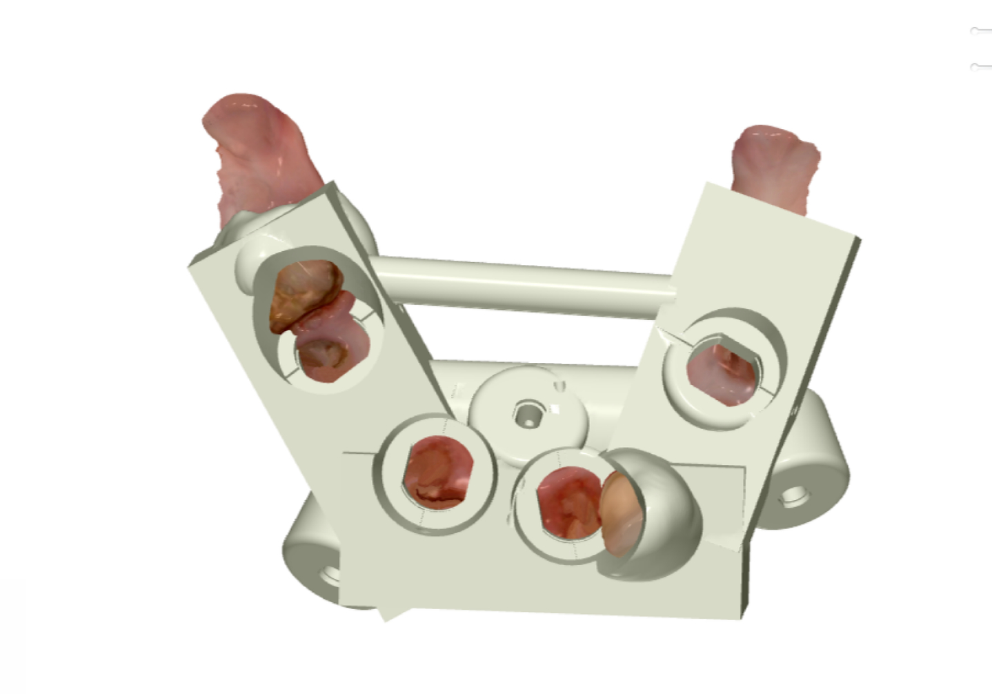

- Manufacturing of Upper Surgical Guides
Phase 3 – Surgical Treatment
- Atraumatic extraction of all remaining teeth
- Guided placement of four Neobiotech IS III Active implants in the maxilla
- Guided placement of four Neobiotech IS III Active implants in the mandible
- Digital implant impression
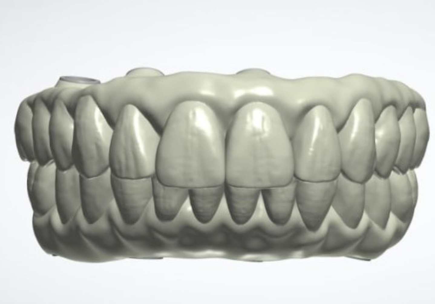
Phase 4 – Immediate Loading
- Fabrication of provisional fixed implant-supported bridges
- Delivery of screw-retained provisional prostheses on the day following surgery
- Occlusal assessment, panoramic radiograph (OPG), and photographic documentation

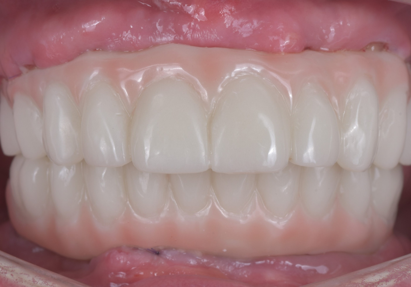


Phase 5 – Follow-up Examination
- Three-month postoperative review
- Evaluation of implant stability, peri-implant soft tissues, and oral hygiene
Phases 6 and 7 – Definitive Prosthetic Rehabilitation
- Removal of provisional bridges and Replacement of Multi-unit abutments
- Definitive digital impressions
- Fabrication of definitive screw-retained prostheses from IPS e.max ZirCAD Prime zirconia supported by individually milled titanium bars
- Delivery of definitive restorations
- Occlusal analysis using the OccluSense® digital system, panoramic radiograph, photographic documentation, and final oral hygiene instruction
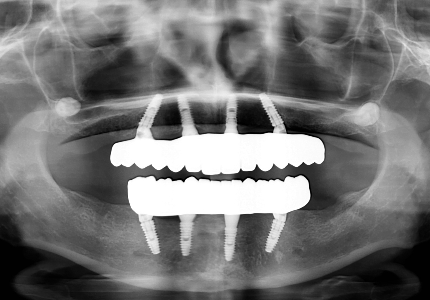


ALL ON 4
Course of Treatment
First Visit – Digital Diagnostic Examination (Approx. 2 Hours)
The initial appointment lasted approximately two hours and focused on comprehensive digital diagnostics.
The examination included:
- Panoramic radiography (OPG)
- CBCT (3D imaging)
- Complete clinical photographic documentation
- Digital intraoral scanning of both the maxillary and mandibular arches
CBCT imaging enabled precise evaluation of bone quality and volume, anatomical structures, and implant treatment possibilities.
Digital intraoral scanning accurately captured the patient’s existing dentition, occlusion, and soft tissues while eliminating the need for conventional impression materials.
A comprehensive consultation was also conducted during the first visit. The clinician thoroughly explained every stage of treatment, the anticipated treatment timeline, and the expected functional and aesthetic outcomes.
Based on all collected diagnostic data, an individualized treatment plan was developed, integrating the surgical, prosthetic, and laboratory phases of treatment.
The total duration of rehabilitation was estimated at approximately six months.
Second Visit – Preoperative Preparation (Approx. 2 Hours)
Due to the advanced periodontal disease, stabilization of the oral environment was required before implant surgery.
During the second appointment, comprehensive professional dental cleaning was performed together with laser-assisted soft tissue therapy.
Retained root fragments of teeth #16, #24, and #25 were atraumatically extracted because they represented chronic sources of infection and were considered non-restorable.
Following completion of periodontal therapy and extraction of the retained roots, new digital intraoral scans of both arches were obtained.
The objectives of this phase included:
- Removal of hard and soft dental deposits
- Elimination of chronic infectious foci associated with retained roots
- Reduction of bacterial biofilm
- Suppression of active periodontal inflammation
- Creation of optimal biological conditions for postoperative healing
- Reduction of the risk of postoperative complications
This preoperative hygiene phase represented an essential component of the overall treatment protocol. By significantly reducing the microbial burden within the oral cavity, it established favorable biological conditions for the subsequent surgical intervention.
As a result, atraumatic extraction of all remaining teeth and immediate implant placement could be safely completed during a single surgical procedure, representing one of the key prerequisites for successful rehabilitation using the All-on-4® treatment concept.
Oral Hygiene Instruction
Patient education formed an integral part of this appointment.
The dental hygienist explained the importance of meticulous oral hygiene before and after implant placement.
The patient received detailed instructions regarding proper toothbrushing techniques as well as future maintenance of implant-supported restorations. She was informed that the long-term success of implant therapy depends not only on a precisely performed surgical procedure but also on consistent daily oral hygiene.
Based on the clinical findings, the patient received individualized recommendations for oral hygiene products, including:
- Toothbrush
- Toothpaste
- Interdental brushes
- Single-tuft brush
- Additional hygiene aids specifically designed for implant-supported fixed prostheses
The patient demonstrated excellent motivation and actively cooperated throughout the entire course of treatment.
Digital Treatment Planning (Laboratory Phase – Approx. 8 Hours)
Following completion of the diagnostic phase, the laboratory workflow was initiated. This phase was carried out without the patient’s presence and required approximately eight hours of combined work by the prosthodontist and the dental technician.
Using specialized CAD/CAM software, data obtained from the CBCT scan, digital intraoral scans, clinical photographs, and the analysis of intermaxillary relationships were integrated into a single digital workflow. Based on these data, a virtual reconstruction of the future dentition was created, including the ideal tooth anatomy, dimensions, morphology, occlusion, and smile aesthetics.
Digital and physical models of both dental arches were subsequently fabricated in our in-house dental laboratory, allowing functional and aesthetic verification of the proposed reconstruction before surgery. The patient was also presented with a digital visualization of her future smile.
Fabrication of Guided Surgical Templates (Approx. 2 Hours)
After completion of the digital treatment planning, two patient-specific surgical guides were manufactured.
One guide was designed for the maxilla and the other for the mandible.
Both templates were tooth-supported, ensuring maximum stability throughout the guided surgical procedure.
The digital surgical plan precisely defined:
- Implant positions
- Implant axis
- Insertion depth
- Implant angulation
- Prosthetically driven orientation
Guided implant surgery enabled highly accurate implant placement according to the pre-planned digital workflow while minimizing surgical trauma and significantly increasing overall treatment precision.
Third Visit – Surgical Phase (Approx. 4 Hours)
Following completion of the digital planning and preoperative preparation, the definitive surgical phase of treatment was performed.
The procedure lasted approximately four hours and was carried out under local anesthesia.
At the beginning of surgery, only those teeth that were not required for stabilization of the surgical guides were atraumatically extracted. Preserving the remaining support teeth at this stage allowed both surgical templates to be positioned with maximum precision and stability during guided implant placement.
By combining CBCT imaging, digital intraoral scans, and virtual treatment planning, the digital treatment plan was transferred accurately to the patient’s oral cavity.
Using computer-guided implant surgery, the following implants were placed:
- Four Neobiotech IS III Active implants in the maxilla
- Four Neobiotech IS III Active implants in the mandible
Each implant was inserted precisely into its pre-planned three-dimensional position, with optimal depth, angulation, and prosthetic orientation for the future definitive restoration.
Thanks to meticulous digital planning and accurate surgical execution, excellent primary implant stability was achieved in all eight implants, allowing implementation of the Immediate Loading protocol.
Only after successful placement of all implants were the remaining teeth, previously serving as support for the surgical templates, atraumatically extracted.
This surgical protocol ensured complete stability of the navigation system throughout implant placement and enabled precise execution of the digital treatment plan without compromise.
After implant placement, each implant was carefully evaluated, the surgical field was thoroughly irrigated and inspected, and Multi-unit abutments were installed to support immediate prosthetic rehabilitation.
Finally, a digital implant impression was obtained using an intraoral scanner and immediately transmitted to the in-house dental laboratory for fabrication of provisional fixed restorations.
Thanks to the fully digital workflow, laboratory production commenced immediately after completion of surgery.
Laboratory Fabrication of Provisional Bridges (Approx. 6 Hours)
Immediately following surgery, fabrication of provisional implant-supported fixed bridges began in our in-house CAD/CAM dental laboratory.
The provisional restorations were produced using the digital implant impressions and the previously approved virtual treatment design, preserving the planned vertical dimension of occlusion, tooth anatomy, tooth length, and smile aesthetics.
These provisional bridges served several important purposes:
- Immediate restoration of aesthetics
- Immediate restoration of masticatory function
- Support and shaping of peri-implant soft tissues
- Stabilization of the newly established occlusion
- Functional verification before fabrication of the definitive zirconia prostheses
Fourth Visit – Immediate Loading of Provisional Bridges (Approx. 1 Hour)
The morning following surgery, the patient returned to the clinic for delivery of the screw-retained provisional restorations.
The appointment lasted approximately one hour.
The provisional bridges were securely fixed onto the implants and comprehensively evaluated.
The clinician assessed:
- Passive fit of the restorations
- Stability of the prosthetic framework
- Adaptation to surrounding soft tissues
- Smile aesthetics
- Vertical dimension of occlusion
- Intermaxillary relationships
- Speech articulation
For maximum precision, a digital occlusal analysis was subsequently performed using the OccluSense® system.
Based on the computerized occlusal analysis, only minor adjustments were required to achieve optimal distribution of occlusal forces across all implants.
Following occlusal adjustment, the following examinations were completed:
- Comprehensive photographic documentation
- Control panoramic radiograph (OPG)
Radiographic evaluation confirmed ideal implant positioning as well as accurate seating of both provisional prostheses.
The patient remained clinically stable without active bleeding or postoperative complications.
Already at this first postoperative appointment, she expressed a very high level of satisfaction with the treatment outcome, particularly appreciating:
- The natural anatomy of the new teeth
- Their length and morphology
- The harmonious appearance of her smile
- Improved comfort during speech
Fifth Visit – Three-Month Follow-up (Approx. 30 Minutes)
Approximately three months after implant placement, the patient attended a routine follow-up examination.
The appointment lasted approximately 30 minutes.
The evaluation included:
- Implant stability
- Healing progress
- Peri-implant soft tissues
- Stability of the provisional restorations
- Oral hygiene
- Occlusal function
Clinical examination demonstrated uneventful healing.
All implants exhibited excellent stability.
The peri-implant soft tissues appeared healthy, pink, and free of inflammation.
No pain, swelling, or bleeding was observed.
The patient’s oral hygiene was evaluated as excellent, and she had consistently complied with all postoperative instructions.
She adapted exceptionally well to the new occlusion and reported complete satisfaction with both the function and aesthetics of the provisional restorations.
She was able to eat comfortably without functional limitations.
Based on the successful osseointegration and healing process, the patient was scheduled for the definitive prosthetic phase of treatment.
Sixth Visit – Definitive Prosthetic Phase (Beginning)
Approximately five months after implant placement, the patient returned to begin fabrication of the definitive implant-supported restorations.
The appointment lasted approximately two hours.
Clinical examination confirmed successful osseointegration of all implants. The implants demonstrated complete stability, while the peri-implant soft tissues had healed fully without any signs of inflammation, allowing transition to the definitive prosthetic stage.
Following removal of the provisional restorations, a thorough clinical evaluation of the implants, Multi-unit abutments, and surrounding peri-implant soft tissues was performed.
As complete healing and soft tissue remodeling had occurred during the five-month healing period, the provisional Multi-unit abutments used for the Immediate Loading protocol were replaced with definitive low-profile Multi-unit abutments better suited to the final soft tissue contours.
This replacement provided several important advantages:
- Improved prosthetic emergence profile
- Enhanced aesthetic integration
- More accurate seating of the definitive restorations
- Easier long-term oral hygiene maintenance
Following replacement of the abutments, scan bodies were attached to each implant, and new digital impressions of both arches were captured using an intraoral scanner.
A new digital bite registration and intermaxillary relationship record were also obtained to provide precise information for fabrication of the definitive prostheses.
All digital records were immediately transferred to the clinic’s CAD/CAM laboratory, where production of the definitive restorations commenced.
After digital impression taking, the provisional bridges were reinstalled, allowing the patient to continue normal daily activities comfortably throughout fabrication of the final prostheses.
Laboratory Fabrication of the Definitive Restorations
After completion of the digital impression workflow, fabrication of the definitive implant-supported prostheses began in the clinic’s in-house CAD/CAM laboratory.
Based on the digital design, individually milled titanium bars were first designed and manufactured.
These titanium bars constitute the structural framework of the entire reconstruction and provide:
- Maximum mechanical strength
- Long-term structural stability
- Precise passive fit on the implants
- Optimal distribution of occlusal forces
- Excellent biocompatibility
Following completion of the titanium frameworks, definitive screw-retained monolithic zirconia bridges were fabricated from IPS e.max ZirCAD Prime, a premium multilayer zirconia material, in shade A1.
IPS e.max ZirCAD Prime was selected because of its:
- Exceptional flexural strength
- Long-term wear resistance
- Natural translucency
- Outstanding aesthetic properties
Each tooth was individually designed according to the anatomy previously approved by the patient during the provisional phase.
Following CAD/CAM milling, the restorations were individually characterized, stained, and glazed by hand to achieve the most natural appearance possible.
Seventh Visit – Delivery of the Definitive Zirconia Bridges
Approximately one month after the definitive digital impressions had been obtained, fabrication of the final prosthetic restorations was completed, and the patient returned for insertion.
The appointment lasted approximately two hours.
The provisional bridges were removed, followed by a final assessment of the implants, Multi-unit abutments, and surrounding soft tissues.
The definitive screw-retained zirconia prostheses supported by individually milled titanium bars were then inserted sequentially.
The clinician carefully evaluated:
- Passive fit of the restorations
- Accuracy of the connection to the Multi-unit abutments
- Adaptation to the peri-implant soft tissues
- Stability of the prosthetic framework
- Smile aesthetics
- Individual tooth anatomy
- Incisal edge position
- Tooth length
- Lip support
- Phonetics
- Intermaxillary relationships
The definitive prostheses precisely replicated the anatomy that had been successfully tested during the several-month provisional phase.
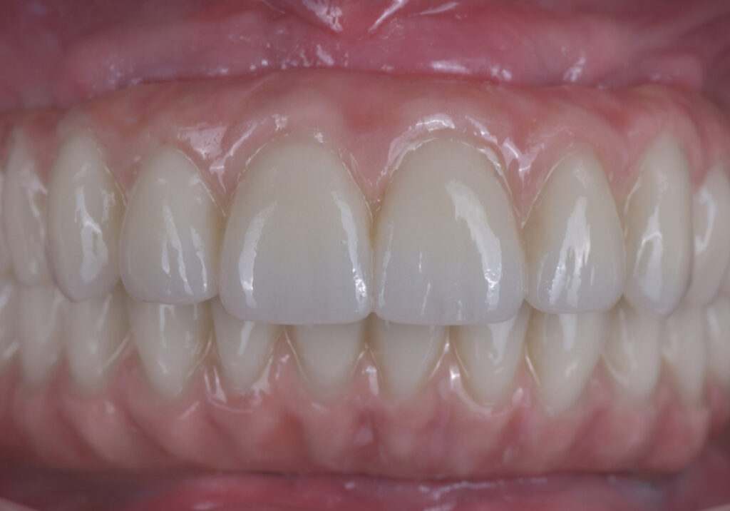
The transition between the restorations and the surrounding soft tissues appeared harmonious and allowed excellent access for daily oral hygiene.
Digital occlusal analysis was again performed using the OccluSense® system.
Only minimal occlusal adjustments were required, resulting in balanced force distribution across the entire prosthetic reconstruction.
Following completion of the prosthetic treatment, the following procedures were performed:
- Panoramic radiographic examination (OPG)
- Complete photographic documentation
- Verification of tightening torque for all prosthetic screws
- Sealing of screw access channels
- Final assessment of function and aesthetics
Radiographic evaluation confirmed accurate seating of both the titanium bars and the definitive zirconia prostheses, as well as ideal positioning of all implants.
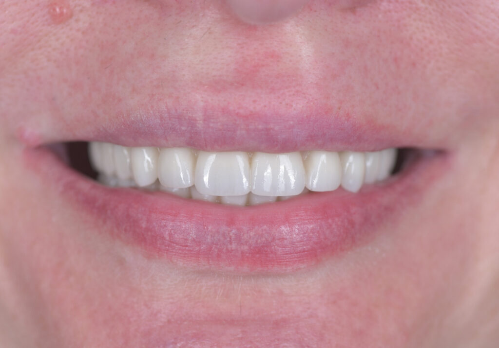
The patient expressed exceptional satisfaction with the final treatment outcome.
She particularly appreciated:
- The natural tooth anatomy
- Tooth length
- Tooth morphology
- Smile aesthetics
- Shade A1
- Comfortable speech
- Comfortable mastication
- The natural feel of her new dentition
Before leaving the clinic, the patient received comprehensive instructions regarding long-term maintenance of implant-supported fixed prostheses.
Proper oral hygiene techniques were reviewed once again, including the use of interdental brushes, a single-tuft brush, and additional implant-specific hygiene aids.
She was enrolled in a regular maintenance program consisting of professional dental hygiene appointments and periodic clinical follow-up examinations, essential for ensuring the long-term success of implant therapy.
Treatment Outcome
The complete oral rehabilitation was successfully completed within approximately six months.
Treatment followed a fully digital workflow integrating advanced diagnostic technologies, guided implant surgery, digital prosthodontics, and in-house CAD/CAM laboratory fabrication.
Complete rehabilitation of both the maxillary and mandibular arches was achieved using the All-on-4® treatment concept.
Thanks to the Immediate Loading protocol, fixed provisional bridges were delivered the day after implant placement, allowing the patient to maintain fixed teeth throughout the entire treatment period without wearing a removable prosthesis.
Following successful osseointegration, the provisional restorations were replaced with definitive screw-retained implant-supported prostheses fabricated on individually milled titanium bars.
The final restorations were manufactured from premium multilayer IPS e.max ZirCAD Prime zirconia in shade A1, combining exceptional mechanical strength, long-term durability, and highly natural aesthetics.
Clinical Results
All planned functional, biological, and aesthetic objectives were successfully achieved.
Treatment resulted in:
- Complete replacement of all hopeless and missing teeth
- Restoration of stable physiological occlusion
- Harmonious intermaxillary relationships
- Natural tooth anatomy
- Ideal tooth proportions
- A highly aesthetic smile consistent with the patient’s expectations
- Individualized shade selection (A1)
- Complete restoration of chewing function
- Significant improvement of phonetics
- Restoration of proper lip support and facial soft tissue contours
Clinical and radiographic follow-up confirmed successful osseointegration of all eight Neobiotech IS III Active implants.
All implants demonstrated excellent stability without mobility or mechanical complications.
The peri-implant tissues healed uneventfully and exhibited healthy pink coloration, firm consistency, and physiological architecture around the definitive prostheses.
No evidence of the following conditions was observed:
- Gingival redness
- Bleeding
- Swelling
- Peri-implant mucositis
- Peri-implantitis
- Implant exposure
- Thread exposure
- Pathological peri-implant bone loss
The transition between the definitive restorations and the surrounding soft tissues remained harmonious while allowing excellent access for daily oral hygiene.
Throughout treatment, the patient demonstrated outstanding compliance with oral hygiene instructions and follow-up recommendations.
Her oral hygiene remained consistently excellent, significantly contributing to uncomplicated healing and the long-term stability of the implant-supported rehabilitation.
Treatment Protocol
- All-on-4® rehabilitation of the maxilla
- All-on-4® rehabilitation of the mandible
- Immediate implant loading
- Screw-retained implant-supported fixed prostheses
- Individually milled titanium bars
- Multilayer IPS e.max ZirCAD Prime zirconia
- Digitally planned guided implant surgery
- Fully digital CAD/CAM workflow
- In-house dental laboratory

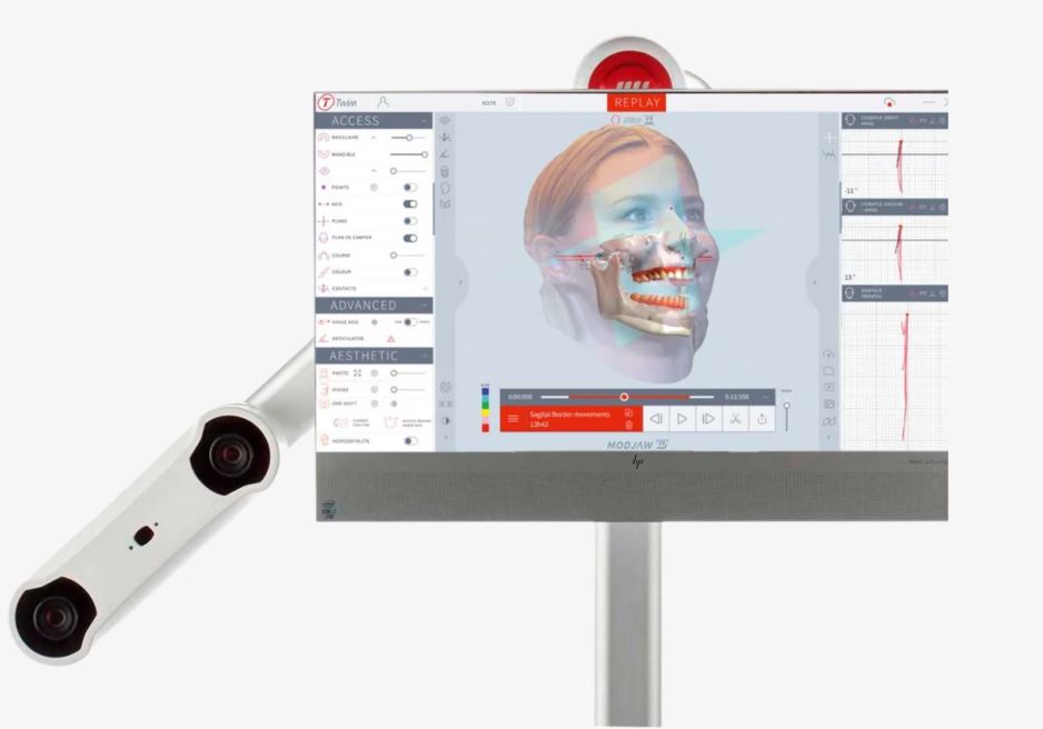


Overall Evaluation
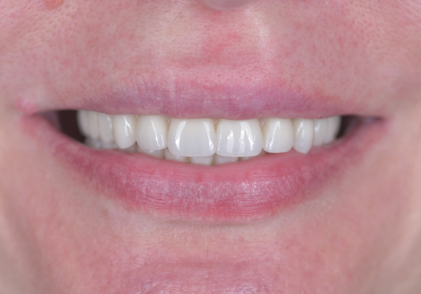
The treatment resulted in a fully functional, biologically stable, and highly aesthetic full-mouth reconstruction that restored the patient’s masticatory efficiency, stable occlusion, comfortable speech, normal eating function, and, most importantly, a natural and confident smile.
The patient expressed complete satisfaction with the final outcome, particularly appreciating the natural appearance of the new teeth, their shape, proportions, color, everyday comfort, and the fact that she never had to wear a removable denture during treatment.
This clinical case demonstrates that the combination of modern digital diagnostics, guided implant surgery, individualized treatment planning, an experienced surgical and prosthetic team, and an integrated CAD/CAM laboratory enables highly predictable long-term outcomes with exceptional functional and aesthetic results.
The All-on-4® treatment concept represents a reliable solution for patients suffering from extensive tooth loss or advanced periodontal disease. When combined with proper case selection, meticulous planning, and regular maintenance care, it provides a durable, stable, and highly comfortable alternative to natural dentition.


Appointment within 24 hours.
We know you don’t want to wait for your health. Fast scheduling for examination and treatment within 2 business days.
Everything under one roof.
No need to go anywhere else – everything is handled in one place with a team of experienced specialists working closely together.
High-quality with warranty.
Top-grade biocompatible materials ensure long-lasting restorations and comfort in everyday use.
Precise 3D diagnostics.
Thanks to advanced 3D technology, we can see every detail, enabling us to plan treatments with maximum accuracy and safety.
"Dental health is not only about technologies – it’s above all about trust, empathy, and collaboration between doctor and patient."